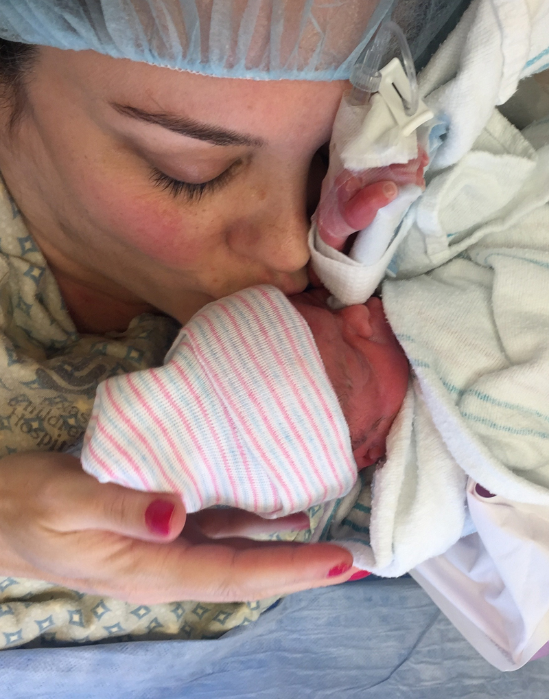
Every first step puts us miles ahead of spina bifida.
We’re here to help parents and children through every step of their journey. That’s why Dr. Michael Belfort and Dr. William Whitehead developed, tested, and proved the fetoscopic spina bifida surgery. This revolutionary procedure reduces developmental issues and drastically improves the chances a child will walk. Because the only way we move forward is by ensuring more first steps.


Every first step puts us miles ahead of spina bifida.
The two-port fetoscopic method was developed to allow for surgery earlier in the pregnancy while reducing risks to the mother found in the open uterus approach. Our revolutionary procedure externalizes the uterus and places just two 4 millimeter ports into the amniotic cavity. Throughout the course of the surgery, we remove most of the amniotic fluid and fill the uterus with carbon dioxide gas, allowing us to visualize the baby via a small camera inserted through one of the ports.
MAKE AN APPOINTMENTBetter outcomes started here
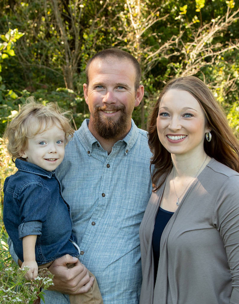
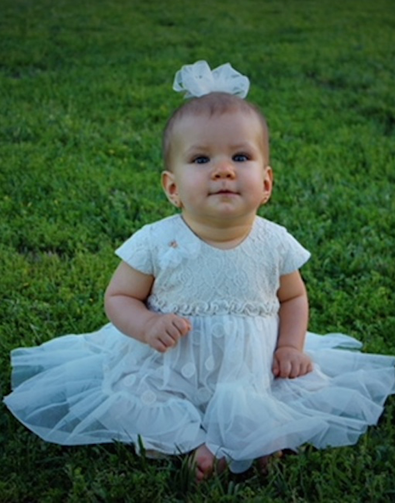
From the Canezaro family to the Camp’s — every smiling face is a gleaming example of our life-changing care.


Minimizing the effects of spina bifida for mothers and babies
Spina bifida occurs when the spine doesn’t close properly during fetal development, causing children to experience a range of physical and mental difficulties. Hospitals around the world treat it using the open-procedure which requires a 7 to 10 centimeter opening made in the uterus, with life-altering risks still present.
The two-port fetoscopic method was developed to allow for surgery earlier in the pregnancy while reducing risks to the mother found in the open uterus approach. Our revolutionary procedure externalizes the uterus and places just two 4 millimeter ports into the amniotic cavity. Throughout the course of the surgery, we remove most of the amniotic fluid and fill the uterus with carbon dioxide gas, allowing us to visualize the baby via a small camera inserted through one of the ports.


 832-822-2229